Research
Development of the Non-contact Measuremet Method of Aspiration Parameters of the Giraffe
Primary function of the lungs is gas exchange, i.e., taking in oxygen and getting rid of carbon dioxide. The lungs can be divided into two parts, alveolus and dead volume. The alveolus is a tiny sac for holding air in the lungs, and dead volume is the part which does not participate in gas exchange. Tidal volume of in humans is smaller than the dead volume, which seems that oxygen does not reach the alveolus and that normal gas exchange cannot continue. However, in fact, axial diffusion is apparently increased by oscillating flow in the airway. This mechanism of gas exchange has been already applied in the field of medical engineering, especially for artificial respirator.
In case giraffe, the ratio of the dead volume to tidal volume is the largest and its aspiratory mechanism is interesting. There are some reports about the aspiration of giraffe, in which respiratory rate, tidal volume and aspiratory flow rate were measured using giraffe with mask. However, the giraffe is one of the most nervous animals in the world, and these reported values may not be correct. Therefore, in this study, an attempt was made to develop a non-contact measurement method of giraffeЃfs aspiration.
A movie of giraffe is shown here. Focusing on the abdominal part of the giraffe, it is confirmed that the abdominal part contracts and expands in response to the aspiration. In this study, we tried to measure aspiratory parameters using this change in shape of the giraffeЃfs abdominal part. Firstly, because the volume changes differently on certain areas of the abdominal part, we decided to divide the abdomen in three, as showm in Fig.1. Red-colored spots are thos of smallest.
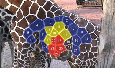
Fig.2 shows area changes of spot # 15, 20, 23, 25 and 26 of the giraffe, and Fig.3 depicts pneumotachogram obtained from Fig.2. From this result, respiratory rate of the giraffe was 10 (breaths/min) and tidal volume was 27 (l). In future studies, we will increase the number of measurements and discuss more precisely.