Brain-Computer Interface
As a communication system for completely locked-in amyotrophic lateral sclerosis (ALS) patients, brain-computer interfaces (BCIs) have received a great deal of attention for several years from a number of research groups. The key technology in a BCI is how to record brain activity and process the obtained data. BCIs are grouped into two categories: invasive and non-invasive. The former, which is also called a brain-machine interface (BMI), requires the implantation of electrodes on or into the cortex. It has the advantage of a relatively high signal noise ratio (SNR) and better spatial resolution. However, the health risks of implantation must always be considered; therefore, non-invasive BCIs are also used.
A non-invasive BCI, such as one that uses electroencephalography (EEG) or near-infrared spectroscopy (NIRS). EEG requires electrodes to be attached to the surface of the head. This type of interface is commonly used to record brain activity indirectly. The problem is that it does not have a good SNR and spatial resolution. Moreover, there are differences in the reproducibility of EEG data among individuals. On the contrary, NRIS accurately reflects brain activity since it can non-invasively measure the blood flow volume and oxygen level of the brain. One of advantage of NIRS is that it is not affected by electromagnetic noise. It is, however, uncertain to what applications it is best suited because it has certain limitations to its practical use. For example, even though the output is only either �gYes�h or �gNo�h, the system still requires a few tens seconds to reach a decision, which is quite a long time.
The ultimate objective of our research is to develop a more practical EEG-based and/or NIRS-based BCI system. To that end, we performed several cognitive experiments to record the brain activity in the frontal cortex while a subject was performing various different tasks. We used the data collected to evaluate the relationship between brain activity and obtained signals.
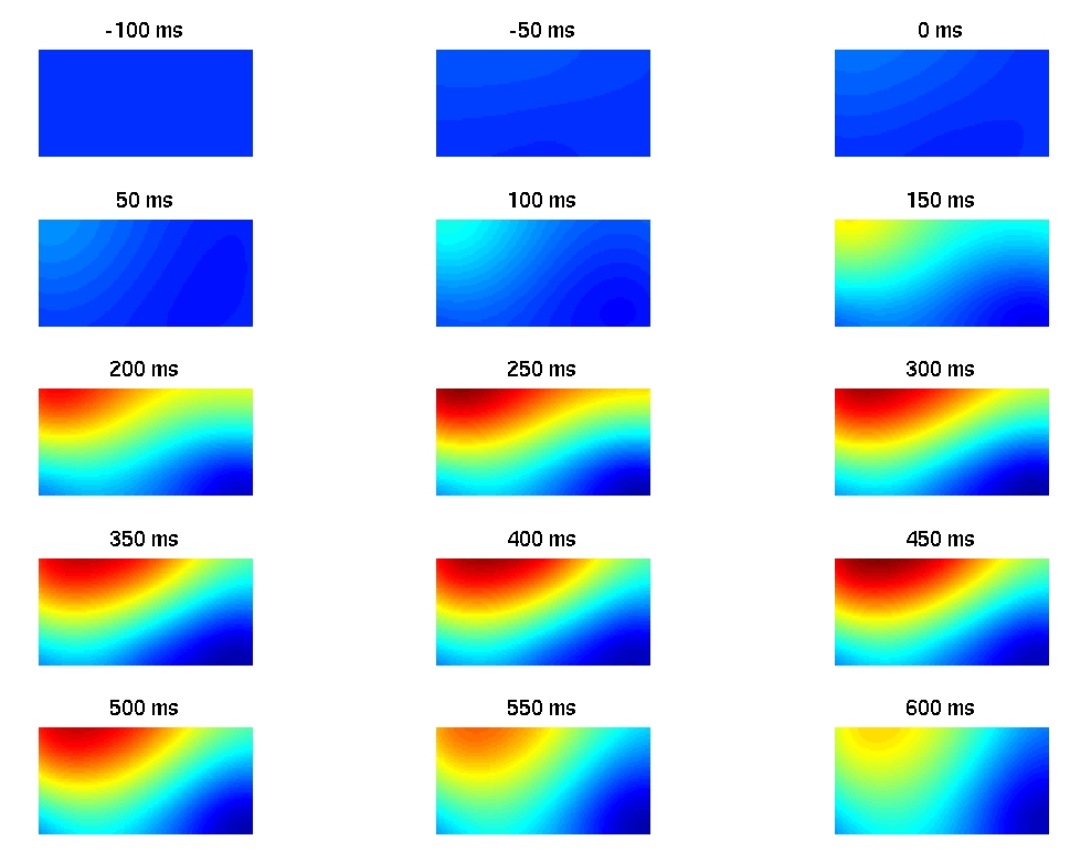
Neural activity in the frontal cortex during a motor imagery task